Blood Vessels Labeled Brain - Blood Vessel Distribution : There is a right sided aca and a left sided aca.. Endothelial cells are labeled in red and pericytes in green. The smallest arterioles consist of endothelium surrounded by a single layer of smooth muscle. Neurosciencenews.com image is adapted from the university of surrey press release. This is particularly important structure due to its clinical implications, which are discussed in more detail in the article. • identification of blood vessels as arteries, capillaries or veins from the structure of their walls.
Blood vessels are referred to collectively as the vascular system and, together with the heart, make up the circulatory system or cardiovascular system. The two cell types ensure the integrity of the neural vasculature by maintaining the blood. Examine a second specimen and notice any differences, such as asymmetries in the size of the vertebral or posterior communicating arteries. The 500 ms patients, both adults and children, also underwent mri scans of the brain to measure iron deposits in surrounding areas of the brain. This is particularly important structure due to its clinical implications, which are discussed in more detail in the article.
Alila Medical Media | Heart and Circulatory System Images from s3.amazonaws.com The 500 ms patients, both adults and children, also underwent mri scans of the brain to measure iron deposits in surrounding areas of the brain. These vessels transport blood cells, nutrients, and oxygen to the tissues of the body. The difference in the structural characteristics of arteries, capillaries and veins is attributable to their respective functions. Endothelial cells are labeled in red and pericytes in green. Most arterioles have all three tunics present in their walls, with considerable smooth muscle in the tunica media. The vessels of the brain circulate blood throughout the brain to ensure that all of its nerves and cells receive the nutrients they need. Blood in the brain is supplied by two pairs of large blood vessels (arteries): • identification of blood vessels as arteries, capillaries or veins from the structure of their walls.
Can you name the main anatomical areas of the brain?.
The arterial blood supply to the brain can be divided into the anterior and posterior circulation. Can you name the main anatomical areas of the brain?. This is particularly important structure due to its clinical implications, which are discussed in more detail in the article. Comes off the subclavian a., ascends although the internal carotid a. The former is derived from the left and right internal carotid arteries, and the latter is haemorrhagic strokes occur when there is a rupture of a blood vessel or abnormal vascular structure within the brain. Supplies the posterior brain, blood supply to the entire brain is ensured by anastomoses between the vessels. They also take waste and carbon dioxide away from the tissues. Blood vessels are referred to collectively as the vascular system and, together with the heart, make up the circulatory system or cardiovascular system. Blood is also supplied to the brain by the vertebral a. Learn the anatomy of a typical human cell. Label the veins of the anterior forearm and hand. The carotid arteries and the vertebral arteries anterior cerebral artery (aca): The vessels allow blood to be pumped at a high pressure to deliver nutrients and.
Endothelial cells are labeled in red and pericytes in green. Identify all of the blood vessels that are illustrated in the figure as you can while holding or otherwise examining whole brain specimens. The former is derived from the left and right internal carotid arteries, and the latter is haemorrhagic strokes occur when there is a rupture of a blood vessel or abnormal vascular structure within the brain. The dense tight junctions between endothelial cells prevent paracellular transport through the. The two cell types ensure the integrity of the neural vasculature by maintaining the blood.
Neuro Anatomy Blood Supply at South College - StudyBlue from classconnection.s3.amazonaws.com Laser ablation was performed on astrocytic endfeet or blood vessels located within a 100 µm depth from the brain surface. The blood vessels are the components of the circulatory system that transport blood throughout the human body. Blood vessels are referred to collectively as the vascular system and, together with the heart, make up the circulatory system or cardiovascular system. Ultrasound may offer a safe way to more as the name suggests, this is a barrier between the brain's blood vessels (capillaries) and the cells and other components that make up brain tissue. The dense tight junctions between endothelial cells prevent paracellular transport through the. Blood is supplied to the brain through 2 major pairs of arteries. Internal carotid artery (anterior circulation), vertebral artery (posterior circulation), and their hexagonal anastomotic network called blood brain barrier refers to the wall between the brain tissue and blood vessels. This vessel supplies blood to the front part of your brain, knows as your frontal lobe.
He says the restricted vessels prevent the blood from draining fast enough and injure the brain by causing a build up of iron which leads to ms.
Traditionally, pais has been explained as being caused by a blood clot forming within the ageing placenta. Microscopically, it is formed by the endothelium of the blood vessel. Endothelial cells are labeled in red and pericytes in green. Neurosciencenews.com image is adapted from the university of surrey press release. This vessel supplies blood to the front part of your brain, knows as your frontal lobe. Laser ablation was performed on astrocytic endfeet or blood vessels located within a 100 µm depth from the brain surface. Towards the anterior side of the brain, those arteries are the internal carotid arteries. Blood vessels are referred to collectively as the vascular system and, together with the heart, make up the circulatory system or cardiovascular system. A laser beam was scanned. This is particularly important structure due to its clinical implications, which are discussed in more detail in the article. Blood travels from the heart in arteries, which branch into smaller and smaller vessels, eventually becoming arterioles. The difference in the structural characteristics of arteries, capillaries and veins is attributable to their respective functions. Blood is supplied to the brain through 2 major pairs of arteries.
Endothelial cells are labeled in red and pericytes in green. Label the veins of the anterior forearm and hand. What we see here are the blood vessels that extend along the inferior surface of the temporal and occipital lobe. Vessel clusters lying within each region were labeled accordingly. Learn the anatomy of a typical human cell.
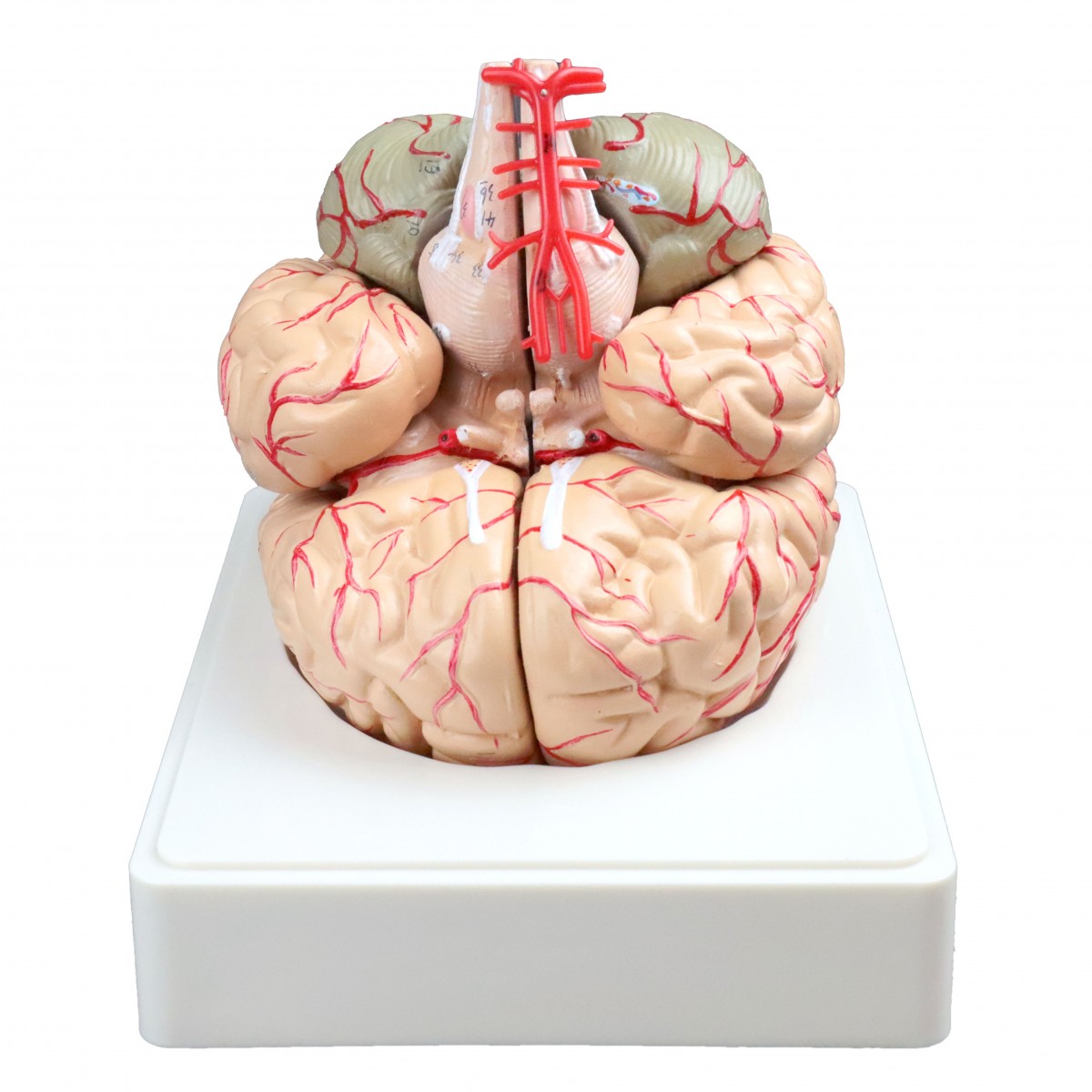
VAB443 Brain with Arteries - Brains & Nervous System ... from www.visionsci.com This vessel supplies blood to the front part of your brain, knows as your frontal lobe. Blood in the brain is supplied by two pairs of large blood vessels (arteries): The smallest arterioles consist of endothelium surrounded by a single layer of smooth muscle. Supplies the posterior brain, blood supply to the entire brain is ensured by anastomoses between the vessels. Label the veins of the anterior forearm and hand. The blood vessels (and nerves) enter the brain through holes in the skull called foramina. The 500 ms patients, both adults and children, also underwent mri scans of the brain to measure iron deposits in surrounding areas of the brain. Most arterioles have all three tunics present in their walls, with considerable smooth muscle in the tunica media.
Function and homeostasis of the brain relies on communication between its complex network of cells.
Learn the anatomy of a typical human cell. The vessels of the brain circulate blood throughout the brain to ensure that all of its nerves and cells receive the nutrients they need. Vessel clusters lying within each region were labeled accordingly. There is a right sided aca and a left sided aca. Label the veins of the anterior forearm and hand. They also take waste and carbon dioxide away from the tissues. The best websites voted by users. The blood vessels are the components of the circulatory system that transport blood throughout the human body. • identification of blood vessels as arteries, capillaries or veins from the structure of their walls. Ultrasound may offer a safe way to more as the name suggests, this is a barrier between the brain's blood vessels (capillaries) and the cells and other components that make up brain tissue. Blood in the brain is supplied by two pairs of large blood vessels (arteries): Blood vessels and lymph nodes. Blood is supplied to the brain through 2 major pairs of arteries.
They also take waste and carbon dioxide away from the tissues blood vessels labeled. Vessel clusters lying within each region were labeled accordingly.
Blood Vessels Labeled Brain - Blood Vessel Distribution : There is a right sided aca and a left sided aca.. There are any Blood Vessels Labeled Brain - Blood Vessel Distribution : There is a right sided aca and a left sided aca. in here.